Blog: Scanning in 7-Tesla: Two years on
In late Spring 2019, UCL’s Queen Square became the setting of an elaborate procedure to install an ultra high-field magnetic resonance imaging scanner to the Wellcome Centre for Human Neuroimaging.
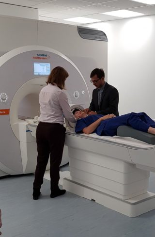
The 19-tonne 7-Tesla MAGNETOM Terra, made by Siemens Healthineers, had to be lowered into the Centre’s specialist scanning suite with a crane.
Now, just over two years later, the Centre’s first studies using the state-of-the-art scanner are beginning to reach completion.
How does a 7T MRI scanner function?
A 7T scanner works largely the same as any other MRI scanner. They produce a strong magnetic field that causes protons inside the body to align, in the same way that a magnet can move the needle of a compass. The scanner then releases short bursts of radio waves, which briefly knock the protons out of alignment. As they realign, radio signals send information about the precise location of protons in the body. Each proton essentially functions as a pixel on a computer screen: when combined in their millions, they build up a detailed image of the brain or other bodily tissues.
But what makes the 7T scanner stand out is the strength of its magnetic field.
“If you go for clinical scan at the hospital, the strength of a normal scan is about 1.5 [Tesla]. Some of them three Tesla,” explains Dr Joost Haarsma, a Research Fellow in the Centre’s Visual Perception team. But with 7T, the magnets are much more powerful. This dramatically increases the resolution of scanning images, revealing more intricate details about the structure of the brain.
“What that allows you to do is essentially to look in finer detail at what is happening in what part of the brain.”
The installation
The arrival of the 7T scanner at the Centre was the culmination of years of hard work. It was part of the Centre’s 2016-2021 funding strategy, for which planning begin in 2014. Centre Director, Professor Cathy Price was a keen proponent of acquiring the high-resolution scanner.
“It was a case of having to secure the funding, which would be the first hurdle,” says Professor Martina Callaghan, Deputy Director and Head of the Physics team at the WCHN, who also played an integral role in getting the 7T scanner to the Centre. “To motivate why it would be a good idea to get the very substantial amount of money that’s involved in installing a system like that. They’re much more complex, from a hardware perspective, so they are more expensive.”
Purchasing the scanner was ultimately made possible by funding from The Leopold Muller fund, Brain Research UK, Wellcome, and UCL’s Capital Equipment Fund. The next steps were procurement, and then preparing to put the system in place – removing an older 3-T scanner in the process.
On the 25th May 2019, the scanner was delivered to the Centre. “A big day for us,” says Professor Callaghan.
Getting this large hunk of metal into the Functional Imaging Laboratory basement proved challenging, and the crane carrying it had to navigate the narrow streets of Queen Square, peppered with obstacles including hotel renovations, street lamps and traffic. Photographs from the installation offer a glimpse into how just major of an operation this was, as well as a rare chance to view Queen Square from above.

For a system this large and complex, Professor Callaghan and the Physics team had to run an extensive series of tests to ensure everything was in working order. In late 2019 the Centre had its first volunteer in the new scanner.
“We were just getting things up and running and beginning to get the studies started. And then the pandemic hit.”
“It was really frustrating and it’s obviously been a lot more difficult, and continues to be difficult to pilot with people,” says Professor Callaghan, citing limited numbers of people allowed on site and struggles to recruit study participants during lockdown as examples of the barriers faced.
But after several long lockdowns and under strict social distancing measures, the Centre’s researchers have finally been able to resume scanning.
New insights into neural structures
For Dr Haarsma, this means utilising the superior resolution of the 7T scanner to explore communication between different layers of the cortex – the outermost layer of the brain, responsible for many higher-order brain functions such as sensation and perception.
“Your grey matter and your cortex is about two and a half millimetres thick. And that’s divided into roughly six layers,” he explains. “In order to look at those individual layers, and how they talk to each other, you need an even smaller resolution. This is what we get with 7T.”
Dr Haarsma’s research is primarily concerned with how the cortex allows us to see things. Specifically, things that aren’t actually there.
“The idea is that the brain has an expectation and these expectations are the brain’s best guess at what’s going to happen next. Now these guesses are often not correct, and when they’re not correct, they need to be corrected.
“If there is a new sensory input, which doesn’t match the expectation, then certain signals in the brain, called ‘prediction error signals’, will correct that expectation. And then hopefully, your best guess is going to be better next time and your perception is correct.”
These ideas fall under the umbrella of predictive coding theories of perception. One theory is that in people who have visual hallucinations, these expectations are particularly strong.
So, where does the 7-Tesla scanner come in?
“These predictive coding theories suggest that it’s particularly the deep layers that become activated by expectations,” says Dr Haarsma, “So expectations flow from higher up in the brain all the way down to the visual parts of the brain. And then sensory input comes in at the middle layers, it gets sent to the superficial layers, and the superficial layer sends it back up.”
If it is true that these expectations are related to the deeper cortex layers, and hallucinations are a result of stronger expectations, then researchers should be able to observe increased activity in the deep cortex when people are hallucinating.
The powerful 7T scanner is needed to distinguish between distorted expectations and another possible culprit for hallucinations: the visual input system.
“You can only tease those things apart when you have a high enough resolution to look at the different layers.”
Incredible detail
And while it’s still early stages and data analysis is ongoing, the images from the scanner are showing a lot of promise.
“What we can say is that we do get different signals from different layers. So it seems to work and we do indeed have a high resolution,” says Dr Haarsma.
Dr Peter Kok, Principal Investigator of the Visual Perception Team, is also pleased with the results so far: “It’s looking really good. We’ve compared the kind of data we’ve been getting from these different scanners and as it stands it’s looking promising.”
“What’s also really crucial for that is the Physics team we have at the [Centre],” he says, “Because this magnet doesn’t do anything on its own. It all depends on programming it in the right way so that you get the right type of data out of it. And that’s what the Physics team is really good at. So we’ve been working with the Physics team and optimising the data collection.”
Joining forces with other groups across the Centre and the wider UCL community has also been one of the highlights for Physics team leader Professor Callaghan:
“What I absolutely love about it is the attitude people have. The people who are most active in the area are just really excellent to collaborate with. They want to do new things and they’re always coming to us with new challenges. I would say that’s invigorating.
“It was really very much a collective effort to get the system to the Centre, and then make the most of the system once it’s at the Centre, despite all of these difficulties.”
When the Centre reflects on the challenges faced, from acquiring funding, to maneuvering a 19 tonne magnet with crane, to the disruption caused by the pandemic: was it all worth it?
“Definitely,” says Professor Callaghan, “When I sit there and the images come in, they just look so good.”
“It’s not all roses – there are harder bits we have to work with. But sometimes you just think ‘Oh, that’s so detailed, that image’, and that’s incredibly rewarding.”