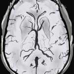
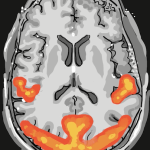
Understanding MRI: Two animations co-created alongside research participants to explain key aspects of magnetic resonance imaging (MRI)
The Understanding MRI team have co-created two new animations which explain important aspects of magnetic resonance imaging (MRI) – a key neuroimaging technique frequently used by researchers at UCL’s Department of Imaging Neuroscience.


Launched this week, the two animations ‘Having an MRI scan for research: What happens?’ and ‘Why do we use MRI for research?’ aim to answer questions that are frequently asked by research volunteers when participating in research at the Department.
Animated by Alice Haskell, the videos set out to explain key aspects of the MRI technology and the experience of having an MRI scan.
Funded by a Departmental Public Engagement grant, the project builds on the success of the team’s first animation ‘What is functional MRI’. The video has had over 22,000 views since its release in 2021.
The project brought together members of several teams across the Department, including the PLORAS, Physics, qMAP-PD and the Imaging Support teams, all of whom work closely with research volunteers. The core team was also supported by a Working Group of researchers from across the Department.
Dr Nadine Graedel and Dr Barbara Dymerska from the team:
“At our Department, we rely on volunteers who are scanned for neuroscience studies and technical research. Often the participants are curious about the inner workings of the MRI scanner, whether it is safe and why we use MRI. We hope that these videos will help address some frequently asked questions, encourage conversations between volunteers, patients, researchers, radiographers, and MRI physicists, and create a positive and constructive experience for all the participants.”
To ensure the animations were accessible and engaging, the team worked closely with research volunteers who had previously participated in research at the Department. Through a series of online focus groups, the volunteers co-designed the animations alongside the team, by shaping the script, narration and imagery of both animations.
Charlotte Dore and Kate Ledingham from the team:
“The best part of the project was involving our research volunteers in the animation development. They were able to bring their first-hand experience of having a research MRI scan to help us shape the videos. Their time and thoughtful input has really improved all aspects of the animations, and we hope that both videos will be viewed by many people who are able to discover more about how MRI works and about the experience of having a scan.”
The final videos will be shared widely, including with new and existing research volunteers before they have an MRI scan.
Once you have watched the animations, you can let us know your thoughts by completing these short feedback surveys.:
· Tell us your thoughts on: Why do we use MRI for research? here.
· Let us know your thoughts on: Having an MRI scan for research: What happens? here.
You can find out more about the project here.